Research Highlights
3D Bioprinting Paves the Way for Fertility Preservation with VitroINK®
VitroINK® provided a xeno-free, structurally supportive bioink that enabled the precise 3D bioprinting of a functional testis model, facilitating cell viability, maturation, and in vitro spermatogenesis research.
Category:
3D Bioprinting
Subcategory/cell type:
Structures Printed
Products
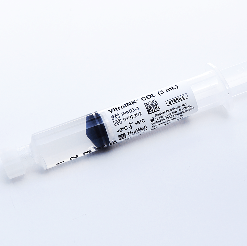
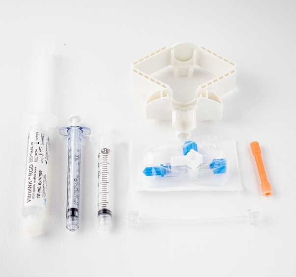
VitroINK® COL (Cat. No: INK03-3)
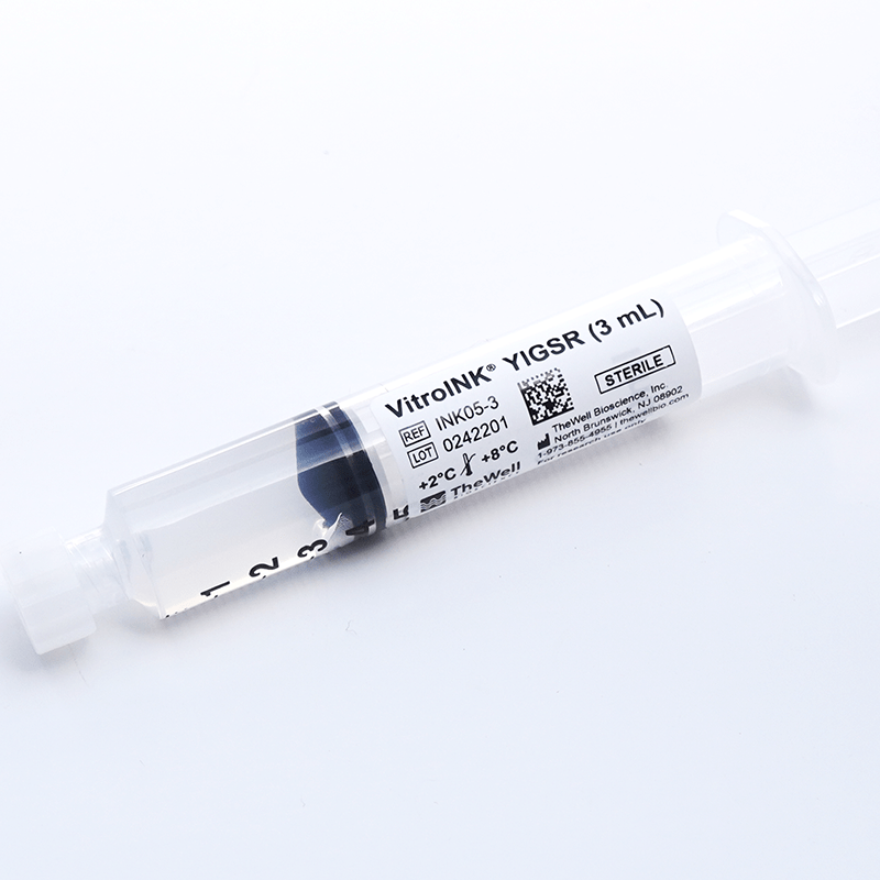
VitroINK® YIGSR (Cat. No: INK05-3)
Cyto3D® Live-Dead Assay Kit (Cat. No: BM01)
Institution:
Vancouver Prostate Centre, Axolotl Biosciences, University of British Columbia, University of Victoria.
Team:
Team: Meghan A Robinson, Sonia HY Kung, Khaled YM Youssef, Kali M Scheck, Robert H Bell, Funda Sar, Anne M Haegert, M Mahdi Asmae, Changfeng Cheng, Salina V Yeack, Bhairvi T Mathur, Feng Jiang, Colin C Collins, Faraz Hach, Stephanie M Willerth, and Ryan K Flannigan
Fertility preservation following pediatric cancer therapy has become a critical area of research, as over 80% of childhood cancer survivors reach adulthood, yet up to 66% of male survivors face infertility due to gonadotoxic treatments. Current fertility preservation methods involve banking testicular tissue in the hope of future in vitro spermatogenesis (IVS) techniques enabling sperm production. However, research in this field has been hindered by the scarcity of human prepubertal testicular tissues and the limitations of animal-based models, which do not fully replicate human spermatogenesis. Additionally, conventional biomaterials used in testis tissue culture often rely on animal-derived components, introducing variability and limiting clinical translation.
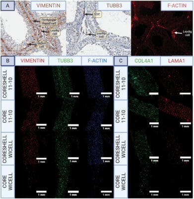
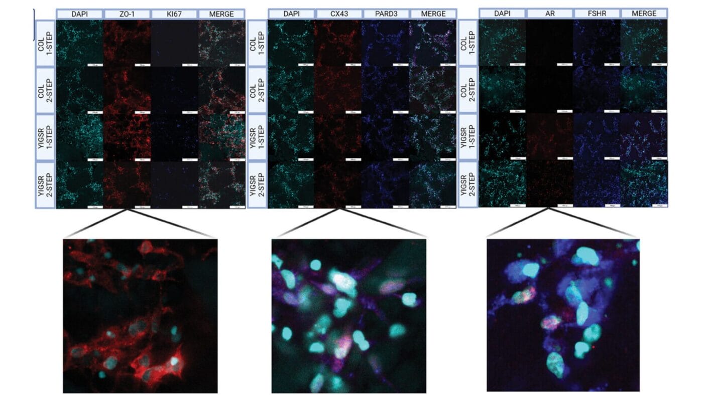
To overcome these challenges, Robinson et al. employed a 3D bioprinted coaxial testis model using human induced pluripotent stem cells (hiPSCs) and the animal-free bioink—VitroINK®. VitroINK® YIGSR and VitroINK® COL were used to encapsulate hiPSC-derived Sertoli, Leydig, peritubular myoid, and spermatogonial stem cells, creating core-shell constructs that mimic testicular cytoarchitecture. VitroINK® bioinks provided a stable and reproducible environment for cellular cross-talk and maturation, eliminating the variability associated with animal-derived matrices.
This 3D bioprinted model successfully captured essential aspects of prepubertal testis function, supporting cell differentiation and maturation under pubertal-like conditions. Furthermore, the xeno-free platform enabled a comparative study of retinoic acid (RA) supplementation methods, revealing that microsphere-released RA significantly improved testicular activity compared to traditional RA medium supplementation.
By integrating VitroINK®, this study has established a scalable, animal-free, and clinically relevant approach for IVS research, bridging the gap advancement paves the way for future fertility preservation solutions, offering new hope for childhood cancer survivors facing infertility.