News
Scaling up tissue engineering with 3D bioprinting
(Nanowerk News) A team at the Wyss Institute for Biologically Inspired Engineering at Harvard University and the Harvard John A. Paulson School for Engineering and Applied Sciences (SEAS) has invented a method for 3D bioprinting thick vascularized tissue constructs composed of human stem cells, extracellular matrix, and circulatory channels lined with endothelial blood vessel cells. The resulting network of vasculature contained within these deep tissues enables fluids, nutrients and cell growth factors to be controllably perfused uniformly throughout the tissue.
The team reported the creation of thick human tissues (>1 cm) replete with an engineered extracellular matrix, embedded vasculature, and multiple cell types by using a multimaterial 3D bioprinting method. These 3D vascularized tissues can be actively perfused with growth factors for long durations (>6 wk) to promote differentiation of human mesenchymal stem cells toward an osteogenic lineage in situ. The ability to construct and perfuse 3D tissues that integrate parenchyma, stroma, and endothelium is a foundational step toward creating human tissues for ex vivo and in vivo applications.

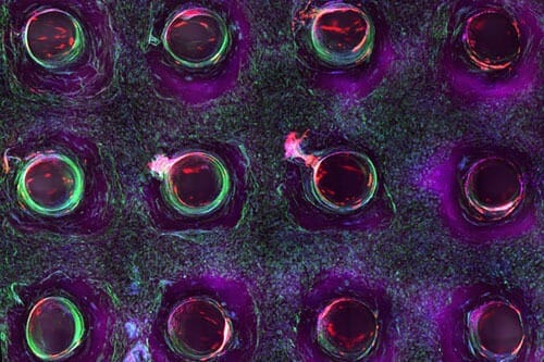
Confocal microscopy image showing a cross-section of a 3D-printed, 1-centimeter-thick vascularized tissue construct showing stem cell differentiation towards development of bone cells, following one month of active perfusion of fluids, nutrients, and cell growth factors. The structure was fabricated using a novel 3D bioprinting strategy invented by Jennifer Lewis and her team at the Wyss Institute and Harvard SEAS. (Image: Lewis Lab, Wyss Institute at Harvard University)
Read more about the report from nano werk: click here
Read more about the article: Three-dimensional bioprinting of thick vascularized tissues

