Research Highlights
Delivery of Cannabidiol via Extracellular Vesicles Improves the Cytotoxic Effects of Chemotherapy on Triple Negative Breast Cancer Cells

Institution:
Florida State University
Team:
Patel N., Kommineni N., Surapaneni S.K., Kalvala A., Yuan X., Gebeyehu A., Arthur P., Duke L.C., York S.B., Bagde A., Meckes D.G., Jr., and Singh, M.
Application:
Creation of a 3D cell culture microenvironment for the delivery of CBD to breast cancer cells via extracellular vesicles.
Disease Model:
Breast Cancer
Hydrogel:
VitroGel® LDP2
Chemotoxic agents such as doxorubicin have been widely employed against triple-negative breast cancer (TNBC), but the method of delivery is critical for maximal effect. Extracellular vesicles (EVs) are being increasingly used for this purpose because, compared to liposomes, they are not as easily recognized by the body as foreign and targeted for clearance. The current study examined the potential for EVs to deliver cannabidiol (CBD) to TNBC cells to increase their sensitivity to doxorubicin. In recent years it has been noticed that CBD may have a positive role in modulating cancer cell proliferation and metastasis, but oral delivery leads to only a small fraction––less than one-fifth of intake––of the compound reaching its target. Thus, the potential for EV-mediated delivery is a promising area for research, and both in vitro and in vivo studies are warranted to examine not only the efficacy of CBD delivery to TNBC cells but also the putative mechanism of how CBD might modulate cancer cell growth and proliferation.
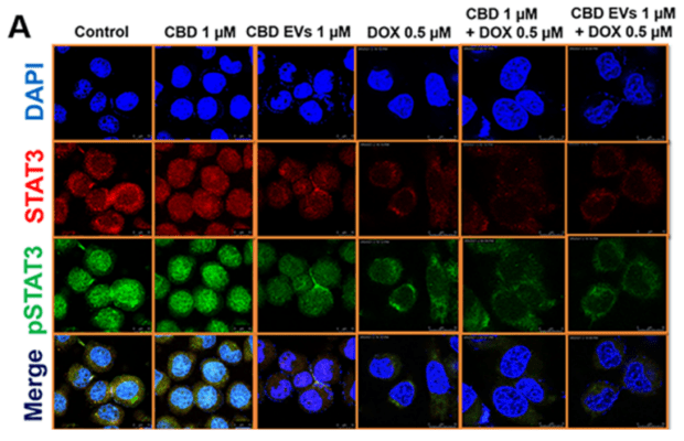
The Florida State team of researchers tested the effects of EV-delivered CBD treatment on TNBC cells in both tissue culture and in a mouse model. In each experiment, controls were included that utilized no CBD, no doxorubicin, and/or no EVs. For in vitro studies, either traditional 2D tissue culture was used, or VitroGel LDP2 was used to create a microenvironment that was a better mimic of live tissue. In either scenario, the cytotoxic effects of CBD and CBD EVs could be followed when incubated with MDA-MB-231 cells after two-day intervals. The EVs were loaded with roughly 10–20% (w/w) with CBD. For in vivo studies, MDA-MB-231 cells were xeno-transplanted into female nude mice and then the animals were treated with CBD, EVs, and doxorubicin under the complete set of control regimens. The subjects were followed for two weeks, with tumor volume measurements and postmortem analyses of tumor cell status. For both 2D and 3D tissue culture studies, CBD delivered via EVs demonstrated significant reductions in the IC50 values compared to CBD or EVs alone. Moreover, cells that had been previously sensitized with 1 mM EV-delivered CBD for one day, followed by a two-day administration of doxorubicin exhibited even further reductions in the IC50 values, especially in 3D cell culture. Examination of the morphology and mitotic status of the cells, combined with the results from the in vivo studies led to the conclusion that EVs facilitate rapid internalization of the CBD cargo into tumor cells, resulting in apoptosis. The research suggests that CBD EVs, even at dosages as small as 5 mg/kg could mitigate the tumor burden of TNBC cells in an animal model.


