Research Highlights
Scaffold-Free 3D Adipose Tissue Organoids: Unlocking Obesity Research with VitroGel® 3D
VitroGel® 3D provided a scaffold-free yet supportive matrix that stabilized adipose organoids for imaging and analysis, enhancing the reliability of in vitro obesity research.
Category:
Downstream analysis / imaging
Subcategory/cell type:
Adipose tissue organoids
Institution:
Bonds Biosystem, SBH Diagnostics, Boston University, Greiner Bio-One North America, Inc
Team:
Rafael Dariolli, Raphael Nir, Tova Mushlam, Glauco R. Souza, Stephen R. Farmer, Miguel L Batista Jr.
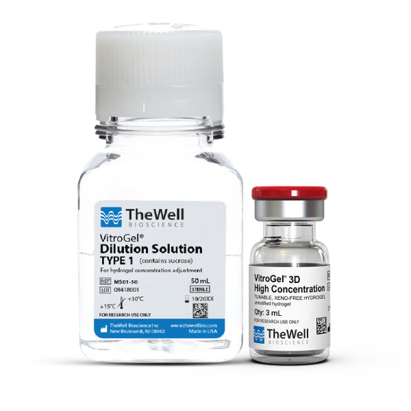
Hydrogel:
VitroGel® 3D (Cat. No: TWG001)
Obesity and metabolic diseases remain urgent global health challenges, yet studying human adipose tissue in vitro is complicated by the limitations of 2D cultures and scaffold-based models. Traditional methods often fail to maintain physiologically relevant adipocyte characteristics, hindering research on adipose tissue function and dysfunction.
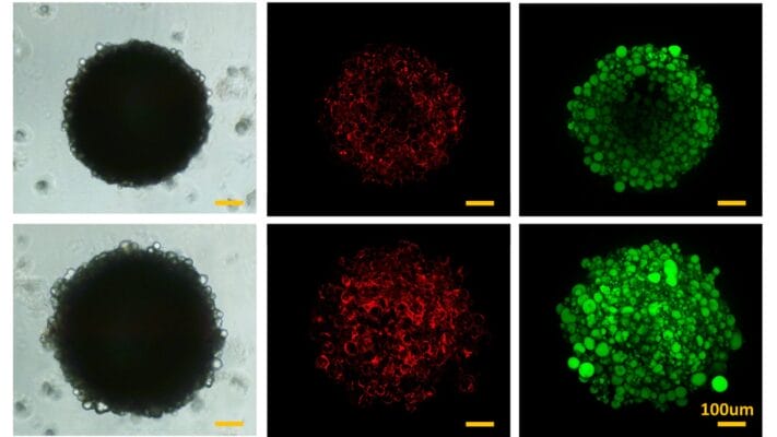
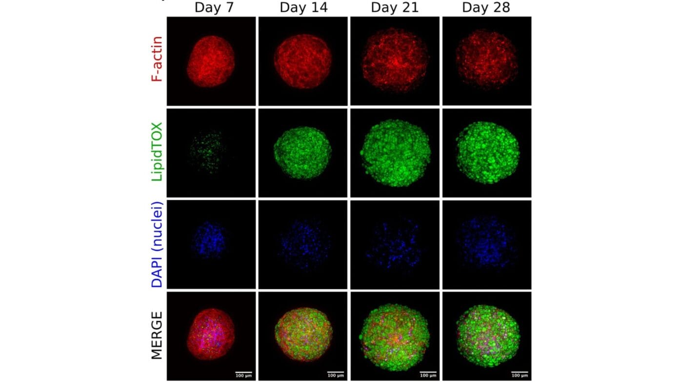
In a recent study, researchers developed a scaffold-free 3D adipose tissue organoid system to better mimic in vivo conditions. To improve imaging and analysis, they embedded the organoids in VitroGel® 3D, ensuring structural stability during immunofluorescence staining and high-content imaging. The unique transparency of VitroGel® enabled high-resolution visualization of adipocyte morphology, lipid accumulation, and cytoskeletal structures without disrupting delicate organoid integrity.
By incorporating VitroGel® 3D, this study enhances the precision and reproducibility of imaging in adipose tissue research, providing a powerful tool for investigating obesity-related metabolic dysfunction and accelerating drug discovery efforts.