Research Highlights
Cells: Prepare Yourself to Make Cartilage

Institutions:
IRCCS Instituto Orthopedico Rizzoli, Bologna, Italy
Team:
Gabusi, E., Lenzi, E., Manferdini, C., Dolzani, P., Columbaro, M., Saleh, Y., and Lisignoli, G.
Disease Model:
Osteoarthritis
Hydrogel:
VitroGel® RGD
Stem cells embedded in VitroGel Hydrogel 3G RGD matrix can display autophagy, allowing a critical examination of this process in the differentiation into healthy chondrocytes.
When a cell starts to chew itself up in order to preserve or recycle its components, the process is termed autophagy. This has proven to be more than desperate attempt to salvage material during times of stress (e.g., starvation); in recent years autophagy is clearly an important homeostatic mechanism that prevents aberrant cellular futures, including arthritis and even cancer. Some key genetic loci control autophagy. These are the autophagy-related genes (ATG) and one of them, ATG5, when disrupted, can trigger events that lead to age-related osteoarthritis (OA). Clearly then, autophagy is an important phenomenon that we need to understand better.
In this paper, a team of immunologists from Italy sought to unravel the cellular events that allow the proper transition from stem cells into chondrocytes, which are cells that make up cartilage. They focused on the molecular genetic changes that occurred during the maturation of adipose-derived mesenchymal stem cells (ASCs) into healthy chondrocytes. Using techniques from molecular biology, immunohistochemistry, and microscopy, they tracked how autophagy related loci change their expression levels as this transformation occurred.
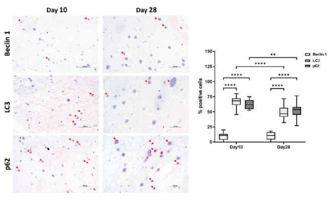
A key aspect of the procedures shown in this paper was the embedding of the ASCs in a 3D hydrogel. The hydrogel provides a microenvironment that seems to favor chondrogenic differentiation, and thus the authors chose to utilize TheWell Bioscience’s VitroGel 3D RGD hydrogel matrix as a medium in which cellular processes could be studied in the laboratory. They had previous success in investigating human ASCs in VitroGel 3D RGD, attributing it to the arginine-glycine-aspartic acid tripeptide motif that this hydrogel motif contains. In this study, the authors embedded 2 x 10⁶ hASCs in 300 μL of a 1:2 dilution of VitroGel 3D RGD, treated them with various growth factors, and tracked the cellular responses over time with the techniques mentioned above. After a few days, for typical ASC cells, they observed differentiation consistent with autophagy, and they detected an upregulation of three gene products characteristic of autotrophy: Beclin 1, LC3, and p62. Later, after about 28 days, in the cells that began to differentiate into chondrocytes, they detected a decrease in the expression of these markers, at the same time they saw typical changes that have been associated with the development of these types of cells.
This study not only shed light on the role of autophagy in maintaining proper cellular development, but it also confirmed the ability of the RGD hydrogel matrix as a means to support autophagic changes. Autophagy appears to be a critical early stage in the proper development of cells into a cartilaginous structure, and this knowledge can lead to therapeutic options for OA and other diseases.


