Research Highlights
Cooking Up Capillaries – Organ-On-A-Chip with HUVECs

Institutions:
National Chung Hsing University, Taiwan
Team:
Lin, Y.-T., Tung Y.-T., Wong, J.-Y., and Wang, G.-J.
Disease Model:
Microvascular System Biomimetics
Hydrogel:
VitroGel® RGD
Researchers find a facile way to create synthetic microscopic vessels in the laboratory utilizing VitroGel RGD.
Biomimetics is the study of how we can create nature-inspired materials for human use. One construct that would be particularly useful in this regard is a synthetic vascular system. The ability to craft microvessels and capillaries in the laboratory would bring large benefits not only for the study and analysis of such structures but it could also help us repair damaged tissues.
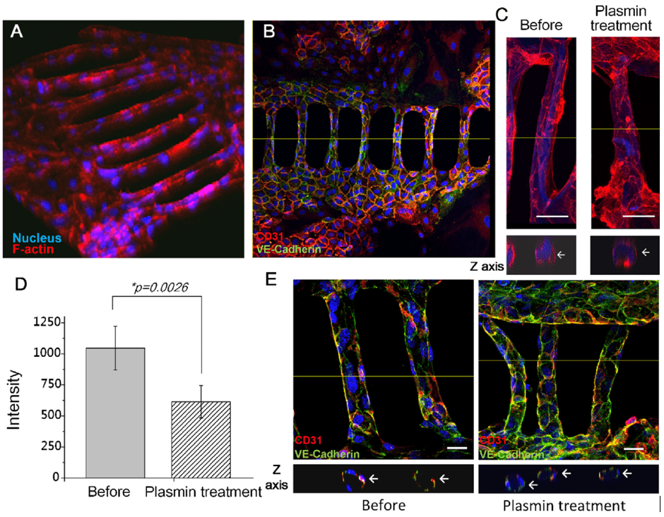
In this paper, a team of Taiwanese mechanical engineers tackled this problem by creating a thin cylindrical scaffold and then growing cultured human cells around it to create a synthetic vessel. The team used photolithography, basically etching with light, applied to polydimethylsiloxane (PDMS) to construct the scaffolds. This was done using microfluidics, or the passage of fluid materials through very thin channels. They then wrapped each scaffold in cultured primary human umbilical vein endothelial cells (HUVECs) to create synthetic vessels. After the degradation of the scaffold using the enzyme plasmin, only the cell-derived structure remained.
An important aspect of this report what that the authors could study the mechanical and biological properties of the synthetic vessels during various stages of preparation. They did this using a series of tests. One was to fluorescently label small molecules (cysteine), which would report on the proper formation of the vascular shape. Another was to examine the structures under a microscope. A third was to test whether the PDMS structures could be successfully perfused with the HUVECs. For this test, the HUVECs were grown in culture for six days and mixed with TheWell Bioscience’s VitroGel® RGD hydrogel. This provided a biological-like medium in which the cells could be evenly distributed throughout the scaffold. Collectively, these tests proved to be successful, and they showed that vessels as small as 30 μm in diameter could be generated, emulating the scale of biological capillaries. These vessels could remain perfusable up to two days after construction.
The methods and data in this paper demonstrated the authors’ ability to fashion a truly biomimetic microvascular system. Because capillary networks are critical to the proper operation of many metabolic functions, and their failure can lead to some serious diseases, the new techniques presented here hold great promise for powerful lab-based experiments that can help us test out potential therapies.


