Research Highlights
Blebs Play an Important Role in Promoting Resistance to Programmed Cell Death in Metastatic Melanoma Cells

Institution:
UT Southwestern Medical Center
Team:
Weems A.D., Welf E.S., Driscoll M.K., Mazloom-Farsibaf H., Chang B.-J., Weiss B.G., Chi J., Dean K.M., Fiolka R., and Danuser, G.
Application:
In vitro encapsulation of melanoma cells either to allow or to block substrate adhesion
Disease Model:
Melanoma
Hydrogel:
VitroGel® RGD and VitroGel® 3D
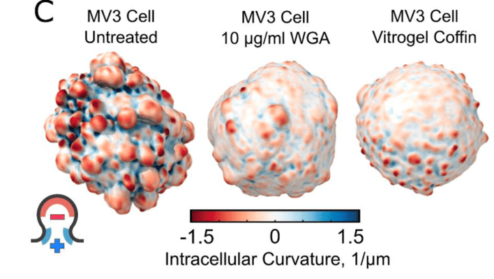
Cancer progression in pre-tumor cells is controlled by a variety of factors. For metastasizing cells, one of these factors is adhesion to a substrate. Cells that cannot anchor to a surface can undergo a form of programmed cell death called anoikis, and thus resistance to this pathway is critical for tumorigenesis. Recently the role of blebs in cell adhesion and anoikis resistance has become appreciated. Blebs are cell protrusions of hemispherical shape that can form during apoptosis, but also during cellular detachment, mitosis, and motility. This latter type of bleb is ephemeral, and if substrate attachment is not made within 1–2 hours, anoikis can result. Cancer cells somehow avoid this fate. This study examined the morphological and associated cellular and molecular changes that occur at various states of blebbiness in order to see if it can be an indicator of the aggressiveness of pre-tumor tissue. The investigators focused on melanoma as a model system but were also interested in the role of blebs in other cancers. The study artificially spurred bleb formation in vitro and then sought correlations with anoikis resistance in melanoma cell lines.
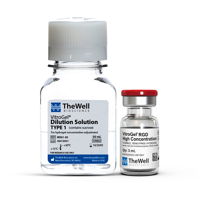
Bleb formation was blocked in MV3 melanoma cells through treatment with wheat germ agglutinin (WGA), which is a lectin having the capacity to impede the deformation of the plasma membrane. Consequently, it became possible to assay the role of bleb formation on anchorage-dependent cell survival. Cells were tracked over a 24-hour period to see how three different melanoma cell lines survived under varying regimes of WGA dosage. These cells were driven to the melanotic state via mutations in the NRAS or BRAF oncogenes. As a control to ensure that bleb formation was the causative agent for survival, parallel groups of cells were encompassed in “coffins” of VitroGel 3D (for detached cell growth) or in coffins of VitroGel RGD, which contains integrin-binding RGD domains that permit cell adhesion. By comparing WGA-sensitive and WGA-insensitive cells, it was found that significantly more MV3 cells underwent anoikis when encapsulated in non-adherent VitroGel coffins. From this, the study could conclude that blebbing contributes to anoikis resistance in melanoma cells. The proposed mechanism for survival is that blebs recruit curvature-sensitive septin proteins to the plasma membrane, and these proteins trigger signaling pathways that drive cell persistence.
Read the publication:
Related Products:


