Webinars
A Quick Organoid Viability Measurement by Using Cyto3D® Live-Dead Assay Kit
ON-DEMAND WEBINAR
PRESENTER

Kalhara Menikdiwela, Ph.D.
TheWell Bioscience
Research Scientist II

WATCH THE WEBINAR
A Quick Organoid Viability Measurement by Using Cyto3D® Live-Dead Assay Kit
Explore the benefits of Cyto3D® Live-Dead Assay Kit in drug discovery studies and numerous other 2D & 3D applications.
Organoids are three-dimensional (3D) and self-organized tissue structures that mimic an organ’s functional and structural features and are becoming increasingly popular among research communities. Although organoid culture techniques and materials have improved tremendously over the past decade, several areas, including cell viability testing, require further improvements. Identifying cell viability (live-dead cells) is one of the primary yet critical requirements in clinical and basic drug discovery studies.
Traditional cell viability assays are primarily designed for 2D cell culture, which poses numerous limitations when applied to 3D systems like organoids. These limitations include uneven staining across wells, limited reagent penetration, variability in staining due to cell heterogeneity, and higher background signals. Such challenges can result in false positive and inaccurate assumptions, making 3D cell viability assessments more complex than standard 2D assays.

Live-dead cell viability images: Intestinal organoids stained with Cyto3D® Live-Dead Assay Kit. Intestinal organoids were cultured in regulated conditions for 5 days.
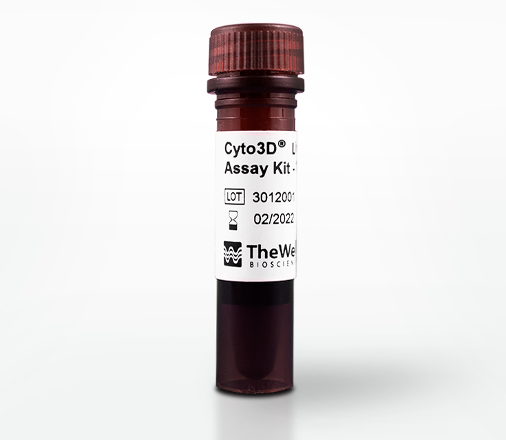
In this webinar, we will demonstrate how to overcome the limitations of 3D cell viability testing with Cyto3D® Live-Dead Assay Kit, a tool designed for both 2D and 3D applications. Cyto3D® Live-Dead Assay Kits can uniformly stain wells with no background signal, making its use ideal in 3D cell culture, 3D spheroid invasion assays, long-term 3D tumor spheroid cultures, hPSCs expansion studies, and 3D bioprinting applications. Other applications of the Cyto3D® Live-Dead Assay Kit, including live-dead cell counting, flow cytometry, single cell sorting, and regular 2D cells live-dead imaging, will also be discussed.
The Cyto3D® Live-Dead Assay Kit is a powerful tool for various applications in both 3D and 2D in vitro studies. It also provides a versatile solution for your live-dead cell testing needs by overcoming challenges associated with traditional cell viability kits.
Key Takeaways:
- Label live-dead cells effectively in 3D tissue structures such as organoids and spheroids.
- Create uniform staining without background noise for clean, clear images with improved resolution.
- Implement a fast, one-step staining procedure that requires NO premixing to cover a broader range of applications in 3D and 2D cell cultures.
PRESENTER
Dr. Kalhara Menikdiwela is a Research Scientist II at TheWell Bioscience. Kalhara earned his doctorate from Texas Tech University, where he investigated the mechanisms linking adipose renin angiotensin system to inflammation and endoplasmic reticulum stress.
As a post-doctoral researcher at the Rutgers University New Brunswick, Kalhara explored the novel role of intestinal fatty acid binding proteins in regulating glucose metabolism and energy homeostasis through specific ligand binding using numerous models including 3D organoids.
At TheWell Bioscience, he oversees the 3D organoid modeling unit primarily focusing on developing and refining various VitroGel® hydrogel systems to support organoid cultures.


